ThinPrep Integrated Imager
MPIN: MP41095
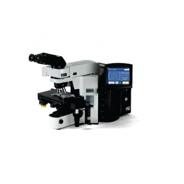
Sign in to view priceThinPrep® Integrated ImagerIntegrates Imaging & slide reviewCombines proven ThinPrep imaging technology and slide reviewing into one system.
Ask for Quote
ThinPrep Integrated ImagerIntegrates Imaging & slide reviewCombines proven ThinPrep imaging technology and slide reviewing into one system.
Benefits
- The review scope can be used as a conventional microscope saving valuable bench space
- User-friendly touch screen and ergonomic stage and review controls
- Automated fiducial mark alignment saves time and increases accuracy
- Automated posts perform can functional checks ensure integrity of data
Features
- Imaging
- Each slide is imaged in approximately 90 seconds
- Every cell and cell cluster on the slide is scanned
- Coordinates of the top 22 largest and darkest objects are identified and stored
Review
- Behaves as an automated microscope, presenting the 22 fields of view (FOVs)
- Dual review process combines human expertise with the power of computer imaging to deliver a superior solution for your lab
- If abnormal cells are identified, the entire slide must be reviewed
- If the 22FOVs are normal, slide can be signed out as negative
- By concentrating on abnormal cases, the cytotechnologist (CT) can screen more efficiently
Shipping Policy
Orders made at Medpick are initiated and processed for shipment upon receipt of request from the customer. Please note that our Shipping Services (Fee, Transportation, Loss or Damage of any shipment, etc.) are in accordance with the Seller\'s terms of Shipment.
Refund Policy
Please refer to Medpick Return Policy.
Cancellation / Return / Exchange Policy
Please refer to Medpick Return Policy.
 REGISTER
 SIGN IN









 Free Shipping

