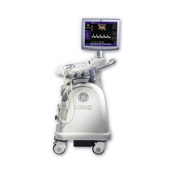
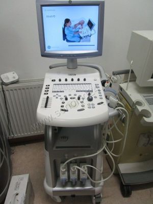
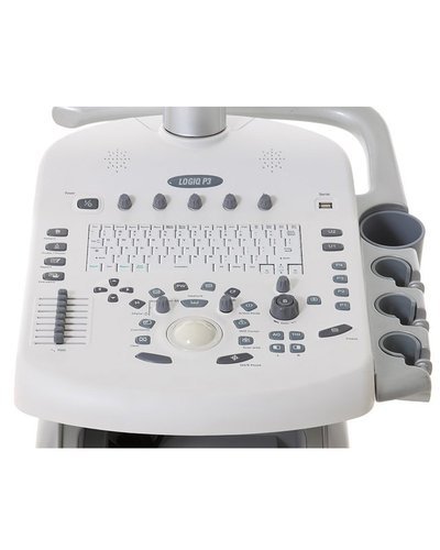
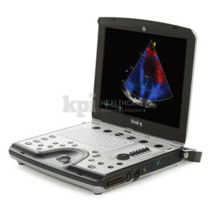
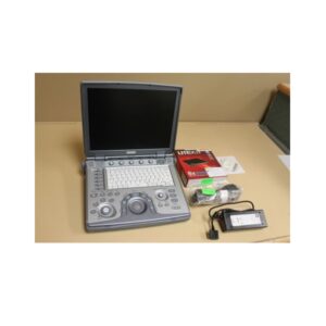
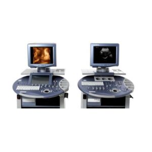
GE Vivid P3 Cardiovascular Ultrasound System
MPIN: MP22618
Sign in to view priceAsk for Quote
GE Vivid P3 Cardiovascular Ultrasound System
The GE Healthcare Vivid P3 is a high performance imaging system designed for cardiac, abdominal and vascular imaging procedures. The unit is based on GE’s TruScanTM architecture provides exceptional ultrasound performance. Its raw data based processing allows images to be viewed, measured, optimized and analyzed online, review, DICOM storage, record archiving and management.
This is done from on-board high capacity hard disk storage. from the cineloop memory and from stored exams, without losing any of the original image quality. It enables virtual re-scan on exams stored a long time ago. Auto Optimization for Tissue, Color and Spectrum – optimized scanning and consistent image quality with one button touch
- Auto TGC for image uniformity with one button touch
- Speckle Reduction Imaging provides reduction in speckle noise, improvedsignal to noise ratio provides higher spatial resolution and deeper penetration
- Live Anatomical M Mode allows easy to obtain on axis measurements innon-standard planes. Plus M-Mode to help assess LV wall motion in apical planes
- Virtual Apex allows a wider field of view in the near field with phasedarray transducers
- LVO Contrast for enhanced visualization of the LV border to help diagnose abnormalities – plus wall motion analysis and ejection fraction calculation
- Stress echo is based on raw data image acquisition, with flexible templates(includes template editor) for exercise and pharmacological protocols, supported by several assessment tools.
Shipping Policy
Orders made at Medpick are initiated and processed for shipment upon receipt of request from the customer. Please note that our Shipping Services (Fee, Transportation, Loss or Damage of any shipment, etc.) are in accordance with the Seller\'s terms of Shipment.
Refund Policy
Please refer to Medpick Return Policy.
Cancellation / Return / Exchange Policy
Please refer to Medpick Return Policy.
 REGISTER
 SIGN IN

