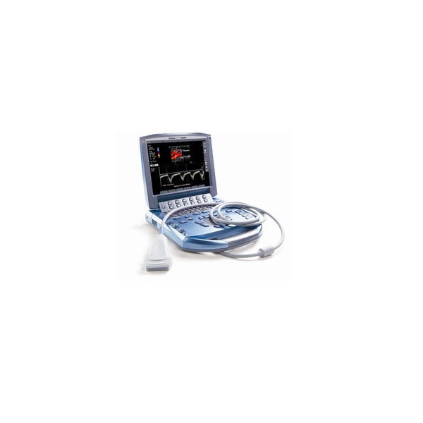
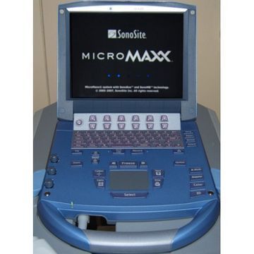
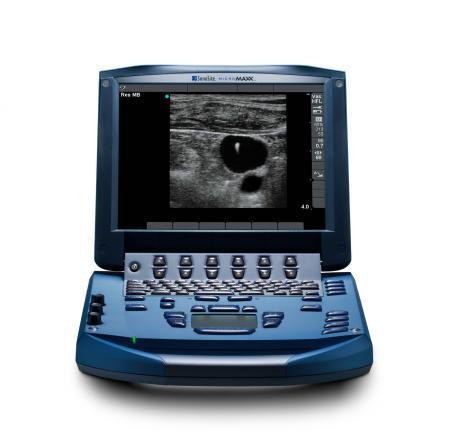
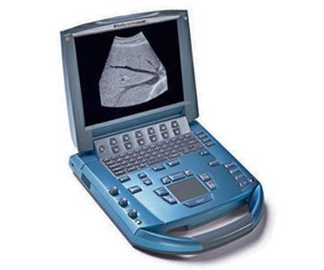
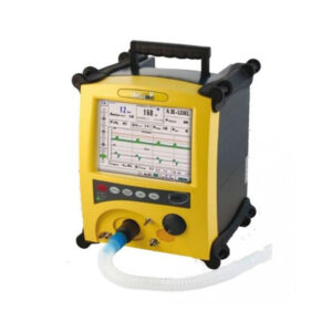
SonoSite MicroMaxx Ultrasound
MPIN: MP22586
Sign in to view priceAsk for Quote
The SonoSite MicroMaxx remains one of our top choices for portable ultrasounds, offering the image quality and portability necessary for a range of applications, from standard point-of-care to emergency. In terms of the SonoSite MicroMaxx price, this portable ultrasound is also one of the least expensive, yet easy and reliable solutions. The MicroMaxx is specifically engineered to take the guesswork out of line placements, cannulation, regional anesthesia, and cardiac assessment, all of which contribute to the safety and well-being of your patients.
Why Choose the SonoSite MicroMaxx Ultrasound
The SonoSite MicroMaxx ultrasound offers several benefits designed to improve machine user confidence, patient safety, and quality of care. Specifically, some of the benefits of the MicroMaxx ultrasound include:
- Portability — Weighing under eight pounds, the MicroMaxx provides premium imaging services without occupying the space in your clinic or hospital.
- Flexibility — In addition to SonoSite’s full cardiac software package and a set of quick-change transducers for anesthesia needs, the MicroMaxx ultrasound has an upgradeable platform that keeps your department ready for expansion.
- Easy to Use — From performance to its intuitive interface, the MicroMaxx is surprisingly easy to use, helping you save valuable time while achieving optimal visualizations of anatomy and needle placements.
- Durability — Including the system design, the results from rugged drop-tests, and its sanitizable keyboard, the MicroMaxx aims to provide peace-of-mind knowing that this ultrasound can survive the fast-paced environments in the OR, the clinic, or the commute in-between.
Imaging Technology
The MicroMaxx ultrasound is a software-controlled system that’s based on an all-digital architecture, using multiple configurations and feature sets to acquire and display high-resolution images. Using 2D, M-Mode, color Doppler, color power Doppler, Tissue Harmonic Imaging, pulsed wave Doppler, continuous wave Doppler, and more, the MicroMaxx’s imaging technologies are engineered to benefit your practice for the following applications:
- Abdominal including the liver, kidneys, pancreas, gallbladder, bile ducts, transplanted organs, and more.
- Cardiac imaging, including the heart, cardiac valves, surrounding anatomical structures, and more.
- Gynecology and infertility imaging, including the uterus, ovaries, adnexa, and surrounding anatomical structures.
- Interventional and intraoperative imaging, including guidance.
- Obstetrical, including fetal anatomy, viability, estimated fetal weight, gestational age, amniotic fluid, and others. CPD and color Doppler is also useful for high-risk pregnant women.
- Pediatric, including abdominal, pelvic, and cardiac anatomy.
- Prostate, including prostate gland.
- Superficial, including breast, thyroid, testicle, lymph nodes, hernias, musculoskeletal structure, soft tissue, and surrounding anatomical.
- Transcranial, including the structures and vascular anatomy of the brain.
- Vascular including the carotid arteries, deep veins, and arteries in the arms and legs; superficial veins in the arms and legs; great vessels in the abdomen, and various small vessels.
Shipping Policy
Orders made at Medpick are initiated and processed for shipment upon receipt of request from the customer. Please note that our Shipping Services (Fee, Transportation, Loss or Damage of any shipment, etc.) are in accordance with the Seller\'s terms of Shipment.
Refund Policy
Please refer to Medpick Return Policy.
Cancellation / Return / Exchange Policy
Please refer to Medpick Return Policy.
 REGISTER
 SIGN IN

