REGISTER
 SIGN IN

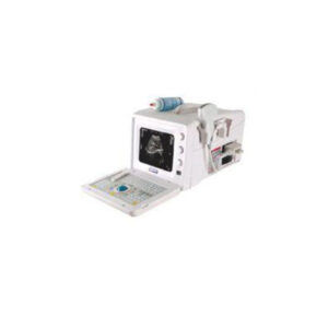
Toshiba Aplio 400 Platinum Ultrasound Machine – Refurbished
MPIN: MP11616
Sign in to view priceAsk for Quote
Review
Toshiba Aplio 400 Platinum Review:
The Toshiba Aplio Platinum series all share the system architecture; therefore, functions in a manner similar to and are intended for the same use. The Toshiba Aplio 400 is a bit lighter than the Aplio 500 because some of options moved, but still the system is overall heavier and larger than newer GE and Philips products. The Toshiba Aplio Platinum series are stable and reliable due to the last 4 to 5 consecutive upgrades since its initial launch in 2012.
The Toshiba Aplio 400 positioned and sold as a high-end system depending on the option configuration, but still holds a very good reputation in Radiology with a wide collection of intraoperative transducers, superb fundamental image quality, and outstanding and dedicated premium radiology options such as Smart Fusion, Shear Wave, and Contrast Harmonic Imaging (CHI).
The Toshiba Aplio Platinum series provides a number of clinical tools that allows for wider clinical utilities and saves scanning time by providing better visualization in a short time. The Superb Micro Vascular Imaging (SMI) is one that increases physician’s clinical productivity and diagnostic efficiency with better visualization of low-velocity blood flow. The MicroPure function is a function that also increases comfort and confidence of diagnosis by better visualizing small structures such as a small calcification in breast. The Toshiba Aplio Platinum series supports unique linear (PLT-308P) and convex (PVT-350BTP) transducers that much improve the needle biopsy procedure by offering the method that a needle can be inserted and visualized in the middle of transducer, not from the edge of a transducer.
The Toshiba products in general look like a technology oriented product, rather than a clinical oriented product. With this series, Toshiba has achieved fully digital beamforming technology, which fully integrated into the Aplio Platinum series. The weight of each Platinum series is still high above 287 lb. The monitor size is 19-inch, which is not compatible with the latest 22 to 23 inches. Although the Toshiba Aplio Platinum holds good quality and reputation in Radiology, they still need to be improved in cardiology and OB/GYN’s workflow, automation, and 4D quality, compared to GE or Samsung products, to satisfy customers.
Probes
Toshiba Aplio 400 Platinum Probes/Transducers:
Convex Probe:
PVT-375BT(6C1)
PVT-375Sc(6CS1)
PVT-674BT(9C3)
Micro Convex Probe:
PVT-382BT(6MC1)
PVT-712BT(11MC4)
Endo Cavity Probe:
PVT-661VT(9C3)
PVT-674BT(10C3)
PVT-781VT(11C3)
PVT-781VTE(11C3)
3/4D Convex Probe:
PVT-375MV(6CV1)
PVT-382MV(6MCV1)
PVT-675MV(8CV2)
PVT-675MVL(9CV2)
PVT-681MV(9CV3)
PVT-681MVL(11CV3)
PLT1204MV(12LV7)
Intra Operative Probe:
PVT-350BTP(6CP1)
PVT-745BTF(11CI4)
PVT-745BTH(11CI4)
PVT-745BTV(11CI4)
PLT-705BTF(11LI4)
PLT-705BTH(11LI4)
Hockey Stick Probe:
PLT1202S(14L7)
BI Plane Probe:
PVL-715RST(11CL4)
Linear Array Probe:
PlT-308P(6LP3)
PLT-604AT(10L4)
PLT-704AT(11L5)
PLT-704SBT(11L4)
PLT-705BT(11L3)
PLT-805AT(12L5)
PLT-1005BT(14L5)
PLT-1204BT(18L7)
PLT-1204BX(18LX7)
Sector Probe:
PST-25BT(5S1)
PST-30BT(5S2)
PST-50BT(6S3)
PST-60ATT(9S4)
TEE
PET-508MA(7S3)
PET510MA(7S3)
PET-510MB(7S3)
PET-511BTM(8SM2)
PET-512MA(8S2)
PET-512MC(8SM2)
PET-512MD(8SM2)
Laparoscopic Probe:
PET-805LA(12LI4)
Pencil Probe:
PC-20M(P2)
PC-50M(P5)
Features
Toshiba Aplio 400 Platinum Features:
- 19-inch wide LCD monitor
- DVD/CD drive
- Precision Imaging
- Tissue Enhancement
- D-THI
- ApliPure+(Spatial Compounding)
- Trapezoid Scan
- Quick Scan
- TDI
- QSP
- HPRF
- Advanced Dynamic FlowTM (ADF)
- DICOM Media Storage
Options
Toshiba Aplio 400 Platinum Options:
Applications
Toshiba Aolio 400 Platinum Applications:
- Abdomen
- Women’s Helacre Care(GYN & Breast)
- Obstetrics
- Fetal Echo
- Vascular
- Transcranial
- Small Parts(Breast, Thyroid, Testis…)
- MSK/Anesthesiology
- Pediatrics
- Urology (Renal, Prostate…)
- Adult Echocardiography
- Pediatric Echocardiography
- Neonate Echocardiography
- Stress Echocardiography
- Adult Transesophageal Echocardiography
- Pediatric Transesophageal Echocardiography
- Internal Medicine w/ Shared Service
- Surgery
- Interventional Radiology
- Contrast Imaging_General Imaging
- Contrast Imaging_Cardiac
- Bowel Imaging
- Strain Elastography
- Shear wave Elastography
FAQs
Toshiba Aplio 400 Platinum FAQs:
• ApliPure reduces ultrasound wave interface within tissues, which appear as speckle patterns or speckle noise on 2D images.
• ApliPure+ can display the boundaries between tissues more clearly and reduce speckle noise and acoustic shadows.
• Precision Imaging allows structures in 2D-mode to be displayed more clearly and the background to be displayed more smoothly.
• Tissue Specific Optimization (TSO) automatically compensates the reception focus.
• Biopsy Enhancement Auto Mode (BEAM) is a function to provide a clearer visualization of biopsy needles in the live ultrasound image. The function provides three enhancement levels and automatically selects the best scan angle.
• 2D Quick Scan enables to automatically optimize the gain and time gain control in 2D mode.
• 2D Wall Motion Tracking semi-automatically extracts wall motion data from the 2D speckle data of the left ventricular myocardial images to be analyzed, and displays the status of myocardial motion.
• Contrast Harmonic Imaging (CHI) is the function that enhances the visualization of vascular mapping with the second-harmonic wave signals from the microbubbles in the contrast medium.
• Parametric MFI allows temporal information to be displayed as a color map superimposed on the CHI-mode image (Contrast image for the period from the start of contrast medium reaches the target region).
• Stress Echo allows the user to perform exercise and pharmacological stress echo exams.
• Luminance is a surface rendering technique that provides a softer, more natural visualization of the human skin resulting in images of photographic impression on quality. The function’s freely movable light source gives the user strong visual feedback on depth and detail. Changing the position of the light can help the user better identify pathological changes and skin defects.
• Fly Thru is the function that enables the user to visualize cavities, ducts and vessels from the inside and in 3D. Comparable to virtual endoscopy, Fly Thru adds cross-sectional ultrasound information to the plain surface data, making it a tool for exploring lesions and ingrowing masses, as well as to assist in planning and follow-up of interventions such as placing stents or grafts.
• Protocol Assistant is a special function that allows for protocols to be created and executed automatically. These protocols can be edited and saves the user time.
• Smart Fusion is the function that allows synchronization of ultrasound scanning with CT/MRI image display. The image is adjusted according to the examination position determined using a magnetic sensor attached to the transducers.
• Smart Navigation is the function that allows a needle navigation line to be superimposed on the ultrasound image based on the positional relationship between the magnetic sensor attached to the transducer and the magnetic sensor attached to the puncture needle.
• Superb Micro Vascular Imaging (SMI) is the function that, with increased Doppler sensitivity, expands the range of visible blood flow and provides visualization of low velocity microvascular flow. SMI’s level of vascular visualization, combined with high frame rates, advances diagnostic confidence when evaluating lesions, cysts and tumors, improving patient outcomes and experience.
• MircroPureTM enables to better visualize small structures in 2D mode. This function shows good image quality for visualizing structures like small calcifications in breast.
• Elastography is the function that enables tissue stiffness to be visualized based on the changes in velocity resulting from physical compression and decompression of the target region.
• Shear wave is the function that allows images representing the speed of propagation of tissue replacement (Shear wave speed) to be visualized (Shear wave scan) by locally displacing tissues by transmitting a burst wave with high acoustic pressure.
• Smart Maps is the function that visualizes and quantifies shear wave propagation in a user-defined region of interest in real time. The user can select both dynamic propagation speed and elasticity displays for visual assessment and quantification.
• Panoramic View allows B/W images to be obtained with a wider field of view by moving the transducer in the lateral direction. Measurement using Panoramic View can be performed.
• FLEX-M is the function that allows any desired plane to be set on both the 2D-mode image and the M-mode image. The set plane can also be reconstructed.
• Auto NT is the function that semi-automatically measures the thickness of nuchal translucency between 12 to 14 weeks in the sagittal section.
Features
Tissue Harmonic Imaging: Yes
Spatial Compounding(=CrossXbeam): Yes(AprliPure+)
Speckle Reduction (=SRI): Yes(Precision Imaging)
Auto Image Opt(B mode) :Yes(Quick Scan)
Auto Image Opt(Doppler): Yes(Doppler scale/Base line)
Write Zoom : Yes
Triplex Mode: Yes
Needle Enhancement or Needle Recognition: Yes(BEAM)
Auto NT Measurement (=Sono NT) : Yes
Auto Follicle 2D Measurement :No
Auto Follicle 3D Measurement :No
Auto IMT : Yes
Auto IMT (Real Time): No
Automated B/M/D Measurement: No
Automated LH Measurement(Automated Function Imaging(AFI), Cardiac Motion Quantification(CMQ), or Auto EF(Ejection Fraction): Yes
Live Dual (B/BC) Mode: Yes
SmartExam or Scan Assistant : Yes(Protocol Assistant)
Fusion : Upgradeable
Raw Data File: Yes
Flexible Report: Yes
Barcode Reader: Yes
Gel Warmer: Yes
Transducers
Convex (1~6Mhz) Yes(6C1)
Convex (2~9Mhz) : Yes(10C3)
Single Crystal Convex (1~6Mhz): Yes(6Cs1)
Single Crystal Convex (2~9Mhz): No
2D Arrary 3D Convex (1~6Mhz): No
Micro Convex (5~8Mhz): Yes(6MC1/11MC4)
Single Crystal Endocavity_Straight Type (3~10Mhz): No
Endocavity_Curved Type (5~8Mhz): Yes(9C3/11C3)
3D Convex (2~6Mhz) : Yes(6CV1/8CV2)
3D Convex Light Weight (2~7Mhz): Yes(9CV2)
3D Endocavity (3~10Mhz): Yes(9CV3/11CV3)
3D Micro Convex (3~9Mhz): Yes(6MCV1)
3D Linear (4~18Mhz): Yes(14LV7)
Linear (>14Mhz): Yes(18L7)
Linear (3~12Mhz): Yes(11L4/11L3)
Linear (<9Mhz): Yes(10L4)
Single Crystal Linear (>14Mhz): Yes(18LX7, Matrix)
Single Crystal Linear (3~12Mhz): Yes(11L4)
Single Crystal Linear (<9Mhz): NO
Linear 50mm : Yes(12L5/14L5)
Linear 25mm : No
Hockey stick (<13Mhz): Yes(14L7)
Hockey stick (>13Mhz): No
T or L shape Intra Operative: Yes(6CP1/11CI4/11LI4)
Phased Array_Adult (1~5Mhz) : Yes(5S1/5S2)
Single Crystal Phased Array_Adult (1~5Mhz) : No
2D Arrary 3D Phased Array (1~5Mhz) : No
Phased Array_Pediatric (3~8hz) : Yes(6S3)
Single Crystal Phased Array_Pediatric (3~8hz): No
Phased Array_Neonate (4~12Mhz) : Yes(9S4)
ICE (Intracardiac Echo Cardiography): No
TEE_Adult (3-7Mhz): Yes(7S3)
TEE_Pediatric (3~7Mhz): Yes(8S2/8SM2)
2D Array 3D TEE (2~7Mhz): No
Pencil CW (2Mhz): Yes(P2)
Pencil CW (5 or 6Mhz): Yes(P5)
Imaging Modes
2D, M mode: Yes
M-color Flow Mode: Yes
Anatomical M-mode: Yes
Trapezoidal Mode : Yes
Color, Power Angio, Pulse Wave Doppler: Yes
Bi-directional Power (=HD FLOW): Yes
SCW Doppler: Yes
Tissue Doppler(Velocity) Imaging: Yes
Freehand 3D: Yes
Live 3/4D OB/GYN: Yes
HD Live: Yes(Luminace)
STIC (Spatio-Temporal Image Correlation): Yes
Live 3D Echo: No
Stress Echo: Yes
Strain and Strain Rate (Cardiac): Yes
B Flow: No
Panoramic Imaging (=Logiq view): Yes
Contrast Imaging – Cardiac: Yes
Contrast Imaging – General Imaging: Yes
Strain-based Elastography : Yes
Shear Wave Elastography: Yes
Applications
Abdominal: Yes
Women’s Health Care (GYN & Breast): Yes
OB : Yes
Fetal Echo: Yes
Vascular: Yes
TCD(Transcranial): Yes
Small Parts (Breast, Thyroid, Testis…): Yes
MSK/Anesthesiology :Yes
Pediatrics: Yes
Urology (Renal, Prostate…) : Yes
Echocardiography_Adult: Yes
Interventional Cardiology: No
Echocardiography_Pediatric: Yes
Echocardiography_Neonate: No
Stress Echocardiography: Yes
Transesophageal Echo_Adult: Yes
Transesophageal Echo_Pediatric : No
Internal Medicine w/ Shared Service: Yes
Surgury: No
Interventional Radiology: yes
Contrast Imaging _ General Imaging (Low MI) : Yes
Contrast Imaging _ Cardiac (High or Low MI): No
Bowel Imaging: Yes
Strain Elastography: Yes
Shear Wave Elastography: Yes
Specification
System Overview
Year Launched: 2014
Estimated Market Price ($): High
Monitor (inch): 19″ LCD
Tilt/Rotate Adjustable Monitor: Yes
Monitor Resolution: 1280×1025
Image Size Resolution :
Touch Screen (Inch): 10.4″
Trackball or Trackpad: Trackball
CP Back-Lighting: Yes
Weight: 130Kg (286.6lb)
Probe Ports: 4+1 (Pencil)
Battery :
Boot-Up Time: No
Sleep Mode (Quick Start): Yes
Maximum Depth of Field: 40cm
Minimum Depth of Field: 1cm
Cart (HCU): No
Independent Steer & Lockable Wheels: Yes
Connectivity
DICOM 3.0: Yes
DICOM SR_Cardiac: Yes
DICOM SR_Vascular: Yes
DICOM SR_OB/GYN : Yes
JPEG, WMV, & AVI: Yes
USB :Yes(5)
HDD/SDD : Yes
DVD/CD RW: Yes
Wireless LAN: Yes
Shipping Policy
Orders made at Medpick are initiated and processed for shipment upon receipt of request from the customer. Please note that our Shipping Services (Fee, Transportation, Loss or Damage of any shipment, etc.) are in accordance with the Seller\'s terms of Shipment.
Refund Policy
Please refer to Medpick Return Policy.
Cancellation / Return / Exchange Policy
Please refer to Medpick Return Policy.