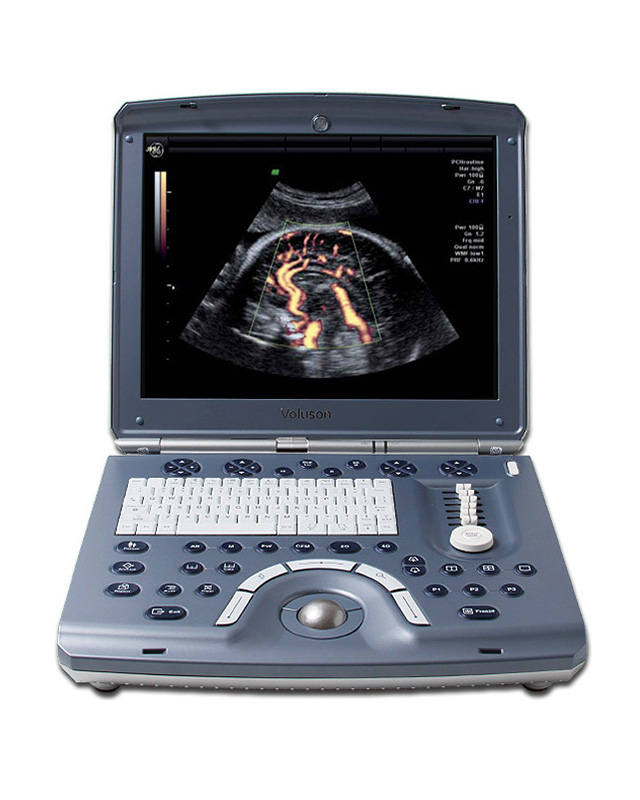
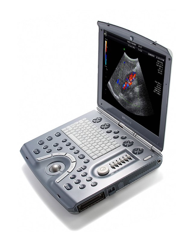
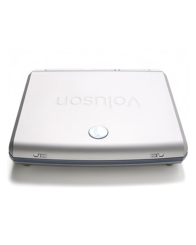
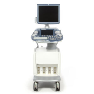
GE Voluson i Ultrasound Machine – Refurbished
MPIN: MP11363
Sign in to view priceAsk for Quote
Overview
Application training for the GE Voluson i
KPI’s on-staff sonographer can provide onsite applications training or remote training via video conference for the Voluson i at a set price plus travel costs. A pre-recorded video training course is included in the sale, lease or rental of the Voluson i from KPI ultrasound.
Voluson i Service options
Free technical support is available from KPI during installation and over the course of the standard limited warranty. Technical support is available after the warranty period at an hourly cost per issue.
GE Voluson i Maintenance
KPI recommends the use of a surge protector along with a dedicated power outlet. Probes should be disinfected after every use with a disinfectant wipe proven not to damage the lens (KPI recommends SonoWipes for this.) KPI recommends one PM (preventative maintenance) every year.
GE Voluson i Dimensions & Weight
Height: 70 mm (2.5 in)
Width: 391 mm (14.2 in)
Depth: 378 mm (12.4 in)
Weight: (no peripherals) 5.7 kg (12.3 lbs.) (includes battery), 5 kg (11 lbs.) (without battery), approx. 38 lbs. with packaging
Voluson i Specifications
Digital Beam former
184.889 MLA2
Displayed Imaging Depth: 0 – 30 cm
Minimum Depth of Field: 0 – 1 cm (Zoom, probe dependent)
Maximum Depth of Field: 0 – 30 cm (probe dependent)
Up to 239 dB Dynamic Range
GE Voluson i Electrical
Voltage: 100 – 250 V
Frequency: 47/63 Hz
Revisions
GE Voluson i Revisions: BT06 to BT07
GE first launched the Voluson i in 2006 at the same time as the Voluson E8. That first version was designated as BT06. “BT” is an abbreviation of “Break Through” and the number designates the year in which this version was launched. So the Voluson i BT06 was launched in 2006 and was in production till the next version in 2007, the v Voluson i BT07. The BT07 revision provided minor bug fixes to the software and support for two new transducers; the RAB4-8-RS 4D high frequency convex and the RNA5-9-RS 4D microconvex.
GE Voluson i Revisions: BT09 to BT14
The Voluson i BT09 revision was a huge update and added the following features and options: VCI, XTD View, STIC, SonoAVC, SonoVCAD Heart, SonoVCAD Labor, HD Flow, and HD-Zoom. In addition to this the GE Voluson-I BT09 also added support for five new transducers: the RIC5-9W-RS 4D intracavitary, the 9L-RS linear, the IC5-9W-RS intracavitary, the AB2-7-RS convex, and the SP10-16-RS high frequency linear. The next revision was BT11 that added the popular SonoNT option to automate the common Nuchal Translucency scan. The current revision in production is the Voluson-i BT14 that adds Volume CINE and support for the RAB2-6-RS 4D convex and 8C-RS microconvex probes. KPI recommends buying the Voluson i BT09 and newer.
All revisions of the GE Voluson i
GE Voluson i (BT06)
GE Voluson i (BT07)
GE Voluson i (BT09)
GE Voluson i (BT11)
GE Voluson i (BT14)
Popular configurations of the Voluson i in 2016
- GE Voluson i BT11 with 2 transducers
RAB4-8-RS 4D Convex
E8C-RS 2D Endovaginal
- GE Voluson i BT14 with 3 transducers
RAB4-8-RS 4D Convex
4C-RS 2D Convex
E8C-RS 2D Endovaginal
Probes
All GE Voluson i probes / transducers
4D Convex RAB2-5-RS [ 1 – 4 MHz ] 192 elements, 46mm, max depth 30cm
4D Convex RAB2-6-RS [ 2 – 6 MHz ] 192 elements, 46mm, max depth 26cm
4D Convex RAB4-8-RS [ 2 – 8 MHz ] 192 elements, 46mm, max depth 26cm
4D Microconvex RNA5-9-RS [ 3 – 9 MHz ] 192 elements, 15.4mm, max depth 18cm
4D Endocavitary RIC5-9-RS [ 4 – 9 MHz ] 192 elements, 11.6mm radius, max depth 16cm
4D Endocavitary RIC5-9W-RS [ 4 – 9 MHz ] 192 elements, 11.3mm radius, max depth 16cm
4D Linear RSP6-16-RS [ 6 – 18 MHz ] 192 elements, 80.7mm volume sweep radius
Convex 4C-RS [ 2 – 5 MHz ] 128 elements, 60.5mm, max depth 30cm
Convex AB2-7-RS [ 2 – 8 MHz ] 192 elements, 41.2mm, max depth 28cm
Microconvex 8C-RS [ 4 – 11 MHz ] 128 elements, 26mm
Endocavitary E8C-RS [ 4 – 10 MHz ] 128 elements, 11.4m, max depth 16cm
Endocavitary IC5-9W-RS [ 3 – 10 MHz ] 192 elements, 11mm radius, max depth 16cm
Linear 12L-RS [ 4 – 12 MHz ] 192 elements, 37mm FOV, max depth 8cm
Linear 9L-RS [ 3 – 8 MHz ] 192 elements, 43mm FOV, max depth 14cm
Linear SP10-16-RS [ 7 – 18 MHz ] 192 elements, 33.7mm FOV, max depth 6cm
Advanced Voluson i probes
The GE Voluson i supports thirteen transducers. As the premier 4D portable the Voluson-i has 6 compatible volume probes including the rare [ 6 – 18 MHz ] RSP6-16-RS high frequency 4D linear transducer excellent for small parts, peripherals, pediatrics and vascular and the RNA5-9-RS 4D microconvex another rare option on any ultrasound machine, especially a portable.
Popular GE Voluson i probes
The RAB4-8-RS is the most popular 4D convex probe with a wide range. The RIC5-9W-RS is the most popular endocavitary probe for the Voluson-i. The 4C-RS is the most popular and inexpensive 2D convex and both the 12L-RS is the most commonly purchased linear probe for the Voluson-i.
Features
GE Voluson i Standard Features
- These are features that are standard on the GE Voluson-i revision BT11.
- 15” TFT LCD Screen
- 1 Active Probe Ports
- 3D/4D Mode
- Innovative user interface with onscreen menus
- HD-Flow
- AO (Automatic Tissue Optimization)
- OTI (Optimized Tissue Imaging)
- Coded Harmonic Imaging
- SRI II (Speckle Reduction Imaging II)
- CrossXBeam CRI
- Static 3D Mode:
- B-Mode only
- B + Power Doppler Mode
- B + CFM Doppler Mode
- B + HD-Flow Mode
- B + CRI
- B + SRI
- B + SRI + CFM
- B + SRI + HD-Flow
- FFC (Focus Frequency Composite)
- Read Zoom
- HD Zoom (High Resolution Zoom)
- Pan Zoom
- Beam Steering
- Virtual Convex
- Beta View
- Trapezoid Mode
- Multi Format (Dual/Quad screen)
- SonoNT (regional availability limitation)
- SonoRenderStart
- Patient information database
- SonoView II: On-board image/data storage software
- CINE mode with CINE Memory: up to 140 MB (3000 2D images)
- Dual/Quad Image
- Review Loop, 4 Review speeds
- Real-Time automatic Doppler Calculations
- Standby Mode
- OB, GYN, Vascular, Neuro, Cardio, Abdominal, Small Parts, Urology, Pediatric
- My Page
- Measurements & Calculations
- Integrated HD
- Proprietary Battery Slot
- Handle
GE Voluson i technology definitions
SRI II:Speckle Reduction Imaging allows the Voluson-i to use a nonlinear diffusion filtering technique that improves image quality in real time by reducing speckles.
CrossXBeam CRI: This is another technology borrowed from the E8. It is compound resolution imaging used to improve border and image clarity the Voluson-i.
FFC: Focus and Frequency Composite is a technology on the GE Voluson i that utilizes two different transmission frequencies and two different focal ranges in the 2D image. This function combines a low frequency to increase the penetration and higher frequency to keep a high resolution. It reduces speckle and artifacts in the 2D image to facilitate the examination of difficult-to-scan patients.
Beta View: On the Voluson-i this feature allows the adjustment of the Volume O-Axis position of 3D probes in 2D mode. The green line in the displayed symbol indicates the position of the acoustic block. + and – define the corresponding sweep directions on the Touch screen.
SonoNT: A Voluson i technology that allows for semi-automatic Nuchal Translucency measurements.
SonoRender Start: A feature on the GE Voluson i that speeds up the acquisition of the fetal face in 4D.
Accessories
GE Voluson i Accessories
- Sony UPD-897MD Digital Black & white thermal printer
- Sony UPD-898MD Digital Black & white thermal printer
- Sony UPX-898MD Digital Black & white thermal printer
- Sony UPD-25MD Digital Color thermal printer
- Mitsubishi P95DW Digital Black & white thermal printer
- Mitsubishi CP30DW Digital Color thermal printer
- Sony DVO-1000 DVD Recorder
- CIVCO disposable biopsy guides (for Convex, Linear and Endo-cavity transducers)
GE Logiq P7 Supplies:
- Aquasonic ultrasound gel
- Sono ultrasound wipes
- Sony UPP-110HG thermal printing paper
- Sony UPC-21L color thermal printing pack
- Mitsubishi CK30L printing paper
- Mitsubishi K95HG high gloss thermal printing paper
GE Voluson i ports
- 1 active transducer port
- 2 USB Ports
- Ethernet LAN port
- 1 PCMCIA Slot
- 1 VGA Out Port
- 1 proprietary Docking Port
Options
GE Voluson i Options
These are features that are not standard on the GE Voluson i BT11, but which can be added to the system for an additional cost or when requested at purchase.
3D/4D Expert
4D View PC Software
DICOM 3
4D Biopsy
VCI (Volume Contrast Imaging)
XTD (Extended View)
TUI (Tomographic Ultrasound Imaging)
SonoVCAD heart
SonoAVC follicle
SonoVCAD labor
STIC (Spatiotemporal Imaging Correlation)
VOCAL II (Virtual Organ Computer-aided Analysis)
Voluson Station
Voluson Dock Cart with integrated power supply and three probe connectors
GoPack carrying case
USB Hub
USB Stick
DVD Drive
External Hard Disk Drive
Footswitch
External Monitor Kit with Isolating Transformer
Bluetooth Printer
GE Voluson i optional technology definitions
VCI: Volume Contrast Imaging on the Voluson-I utilizes 4D transducers to automatically scan multiple adjacent slices and delivers a real-time display of the ROI. This image results from a special rendering mode consisting of texture and transparency information. VCI improves the contrast resolution and therefore facilitates finding diffuse lesions.
TUI: Tomographic Ultrasound Imaging is a new visualization mode for the Voluson-i in 3D and 4D data sets on the Voluson-i. The data is presented as slices through the data set which are parallel to each other. An overview image, which is orthogonal to the parallel slices, shows which parts of the volume are displayed in the parallel planes. This method of visualization is consistent with the way other medical systems such as CT or MRI, present the data to the user. The distance between the different planes can be adjusted to the requirements of the given data set. In addition it is possible to set the number of planes. The planes and the overview image can also be printed to a DICOM printer, for easier comparison of the ultrasound data with CT and/or MRI data.
SonoVCAD Heart: A technology that automatically generates a number of views of the fetal heart to make diagnosis easier.
SonoAVC follicle: A Voluson-i technology that automatically detects follicles in a volume of an organ (e.g., ovary) and analyzes their shape and volume. From the calculated volume an average diameter can be calculated. It also lists objects according to their size.
SonoVCAD labor: Allows the Voluson-i user to measure fetal progression during the second stage of labor such as fetal head progression, rotation and direction. Visual evidence and objective data of the labor process are provided. All SonoVCAD labor measurements are automatically added to the worksheet
STIC: Spatiotemporal Imaging Correlation is a fetal echo provided by the Voluson-i that visualizes the fetal heart or an artery in static 3D.
VOCAL II: Virtual Organ Computer-aided Analysis is an imaging program on the Voluson-i for cancer diagnosis, therapy planning and follow-up therapy control. It offers contour detection of structures and volume calculation. A virtual shell can be set around the contour of the lesion. VOCAL automatically calculates the vascularization within the shell by 3D color histogram by comparing the number of color voxels to the number of grayscale voxels.
Imaging Modes
GE Voluson i Standard Imaging Modes
B-mode (2D)
M-mode (M)
M-Color-Mode (MC)
Color Flow Mode (C)
Power Doppler Imaging (PD)
HD-Flow Imaging (HD-Flow)
PW Doppler / Spectral Doppler Mode (PW)
Extended View (XTD View)
Speckle Reduction Imaging II (SRI II)
CrossXBeam (CRI)
Real-time Triplex Mode
Split, quad screen
Volume Mode (3D/4D) :
3D Static
3D with Color Flow
4D Real-Time
Voluson i Optional Imaging Modes
Volume Contrast Imaging (VCI)
Spatiotemporal Image Correlation (STIC)
Tomographic Ultrasound Imaging (TUI)
Applications
GE Voluson i Applications
Applications or Apps are the types of exams or studies that an ultrasound machine can do. More than this if an ultrasound machine supports a specific application it will have calculations, measurement and reporting software included to support those apps and make them useful in a clinical environment.
The GE Voluson i is able to do a variety of applications but it’s focus is women’s health and 4D.
Obstetrics
Fetal
Gynecology
Abdominal
Fertility
Small-Parts (Breast, Testes, Thyroid, etc.)
Peripheral Vascular
Musculoskeletal conventional and superficial
Transvaginal
Transrectal
Pediatrics
Urology
Oncology
Orthopedics
Vascular
Cardiac (fetal cardio)
Neurology
Specification
System Overview
Year Launched : 2006
Estimated Market Price ($) : High
Monitor (inch) : 15″ TFT LCD
Tilt/Rotate Adjustable Monitor : Yes
Monitor Resolution : 1027*768
Image Size Resolution : 800*600
Touch Screen (Inch) : No
Trackball or Trackpad : Trackball
CP Back-Lighting : Yes
Weight : 12.3lbs(5.6kg)
Probe Ports : 1
Battery : Yes
Boot-Up Time :
Sleep Mode (Quick Start) :
Maximum Depth of Field : 30cm
Minimum Depth of Field : 0-2cm
Cart (HCU) : Yes
Independent Steer & Lockable Wheels : Yes
Imaging Modes
2D, M mode : Yes
M-color Flow Mode : Yes
Anatomical M-mode : No
Trapezoidal Mode : Yes
Color, Power Angio, Pulse Wave Doppler : Yes
Bi-directional Power (=HD FLOW) : Yes
SCW Doppler : No
Tissue Doppler(Velocity) Imaging : No
Freehand 3D : Yes
Live 3/4D OB/GYN : Yes
HD Live : No
STIC (Spatio-Temporal Image Correlation): Yes
Live 3D Echo : No
Stress Echo : No
Strain and Strain Rate (Cardiac) : No
B Flow : No
Panoramic Imaging (=Logiq view) : No
Contrast Imaging – Cardiac : No
Contrast Imaging – General Imaging : No
Strain-based Elastography : No
Shear Wave Elastography : No
Features
Tissue Harmonic Imaging : Yes
Spatial Compounding(=CrossXbeam) : Yes
Speckle Reduction (=SRI) : Yes
Auto Image Opt(B mode) : Yes
Auto Image Opt(Doppler) : Yes
Write Zoom : Yes
Triplex Mode : Yes
Needle Enhancement or Needle Recognition : No
Auto NT Measurement (=Sono NT) : Yes
Auto Follicle 2D Measurement : Yes
Auto Follicle 3D Measurement : Yes
Auto IMT : No
Auto IMT (Real Time) : No
Automated B/M/D Measurement : No
Automated LH Measurement(Automated Function Imaging(AFI), Cardiac Motion Quantification(CMQ), or Auto EF(Ejection Fraction) : No
Live Dual (B/BC) Mode : Yes
SmartExam or Scan Assistant : No
Fusion : No
Raw Data File : No
Flexible Report : No
Barcode Reader : No
Gel Warmer : No
Applications
Abdominal : Yes
Women’s Health Care (GYN & Breast) : Yes
OB : Yes
Fetal Echo : No
Vascular : Yes
TCD(Transcranial) : No
Small Parts (Breast, Thyroid, Testis…) : Yes
MSK/Anesthesiology : Yes
Pediatrics : Yes
Urology (Renal, Prostate…) : Yes
Echocardiography_Adult : No
Interventional Cardiology : No
Echocardiography_Pediatric : No
Echocardiography_Neonate : No
Stress Echocardiography : No
Transesophageal Echo_Adult : No
Transesophageal Echo_Pediatric : No
Internal Medicine w/ Shared Service : Yes
Surgury : No
Interventional Radiology : No
Contrast Imaging _ General Imaging (Low MI) : No
Contrast Imaging _ Cardiac (High or Low MI) : No
Bowel Imaging : No
Strain Elastography : No
Shear Wave Elastography : No
Transducers
Convex (1~6Mhz) : Yes
Convex (2~9Mhz) : No
Single Crystal Convex (1~6Mhz) : No
Single Crystal Convex (2~9Mhz) : No
2D Arrary 3D Convex (1~6Mhz) : No
Micro Convex (5~8Mhz) : No
Single Crystal Endocavity_Straight Type (3~10Mhz) : No
Endocavity_Curved Type (5~8Mhz) : Yes
3D Convex (2~6Mhz) : Yes
3D Convex Light Weight (2~7Mhz) : Yes
3D Endocavity (3~10Mhz) : Yes
3D Micro Convex (3~9Mhz) : Yes
3D Linear (4~18Mhz) : Yes
Linear (>14Mhz) : Yes
Linear (3~12Mhz) : Yes
Linear (<9Mhz) : Yes
Single Crystal Linear (>14Mhz) : No
Single Crystal Linear (3~12Mhz) : No
Single Crystal Linear (<9Mhz) : No
Linear 50mm : No
Linear 25mm : No
Hockey stick (<13Mhz) : No
Hockey stick (>13Mhz) : No
T or L shape Intra Operative : No
Phased Array_Adult (1~5Mhz) : No
Single Crystal Phased Array_Adult (1~5Mhz) : No
2D Arrary 3D Phased Array (1~5Mhz) : No
Phased Array_Pediatric (3~8hz) : No
Single Crystal Phased Array_Pediatric (3~8hz) : No
Phased Array_Neonate (4~12Mhz) : No
ICE (Intracardiac Echo Cardiography) : No
TEE_Adult (3-7Mhz) : No
TEE_Pediatric (3~7Mhz) : No
2D Array 3D TEE (2~7Mhz) : No
Pencil CW (2Mhz) : No
Pencil CW (5 or 6Mhz) : No
Connectivity
DICOM 3.0 : Yes
DICOM SR_Cardiac : No
DICOM SR_Vascular : No
DICOM SR_OB/GYN : Yes
JPEG, WMV, & AVI : Yes
USB : Yes(2)
HDD/SDD : 80GB
DVD/CD RW : Yes(External)
Wireless LAN : Yes
Shipping Policy
Orders made at Medpick are initiated and processed for shipment upon receipt of request from the customer. Please note that our Shipping Services (Fee, Transportation, Loss or Damage of any shipment, etc.) are in accordance with the Seller\'s terms of Shipment.
Refund Policy
Please refer to Medpick Return Policy.
Cancellation / Return / Exchange Policy
Please refer to Medpick Return Policy.
 REGISTER
 SIGN IN

