 REGISTER
 SIGN IN
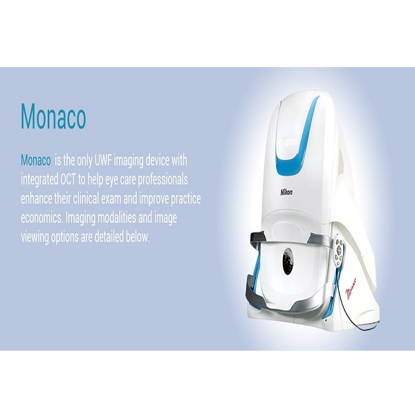
Monaco
MPIN: MP47453
Sign in to view priceMonaco the 200 degree, single capture retinal imaging system with integrated OCT, which shows virtually the entire retina in less than ½ second and provides cross-sectional OCT views.
Ask for Quote
Product Description
Why Monaco?
Monaco the 200 degree, single capture retinal imaging system with integrated OCT, which shows virtually the entire retina in less than ½ second and provides cross-sectional OCT views.
Monaco imaging modalities and image viewing options:
Image Modalities
Color
Red-free
Choroidal
Autofluorescence
OCT
Image Views
Standard: 200⁰ Single Capture
Auto-montage: Up to 220⁰
Central Pole: Detailed view of the macula
Stereo: Image pairing for optic disc and retinal evaluation
OCT: Cross-sectional imaging of ocular structures, including the fundus
Technical Specifications
Image Modalities:
optomap color and optomap plus (red and green laser):
Color composite view
Green laser view
Red laser view
optomap af (green laser): autofluorescence
Optical Coherence Tomography (OCT)
Resolution
optomap color: 20 μm
optomap plus, af : 14 μm
Wavelengths
Red laser: 635 nm
green laser: 532 nm (for af )
Exposure Time
Less than 0.4 seconds
Tomographic Imaging
Signal Type: Optical scattering from tissue
Signal Source: Super luminescent Diode (SlD) 830 nm
Optical Power: laser safety Class-1 following IEC/en60825-1:2014
Typical Axial Resolution: <10 micron (in tissue) Digital on-screen <6 micron
Transverse Resolution: 20 micron (in tissue)
Scanners: Galavanometric with x, y mirrors
Scan Depth: Up to 2.5mm
OCT Scan Characteristics
Spectral Domain OCT
A-Scan rate up to 70k cycles/s
Active eye tracking
Automatic scan positioning
OCT Scan Types
Line Scan
Raster Scan
Retina Topography Scan
Optic nerve Head (OnH) Topography Scan
Retinal nerve Fiber layer (RnFl) Scan
Footprint
Width: 550 mm/22 inches
Depth: 500 mm/19.5 inches
Height: 650 mm/25.5 inches

optomap color
Red Free
Choroidal
Optomap af
OCT
Shipping Policy
Orders made at Medpick are initiated and processed for shipment upon receipt of request from the customer. Please note that our Shipping Services (Fee, Transportation, Loss or Damage of any shipment, etc.) are in accordance with the Seller\'s terms of Shipment.
Refund Policy
Please refer to Medpick Return Policy.
Cancellation / Return / Exchange Policy
Please refer to Medpick Return Policy.
Related products
-
Consumer Goods, Health Innovations, Home Health and Home use innovations, Masks, Personal Hygiene
Smart Air Pollution Mask
Consumer Goods, Health Innovations, Home Health and Home use innovations, Masks, Personal HygieneSmart Air Pollution Mask
The World’s Best Smart Air Pollution Mask
- Inhale across the filter like you don’t wear a mask with the breakthrough BreatheEase™ System
- No Inward leakage of bad air or sore skin with the innovative Sealing System
- Reduced heat and Humidity in the inner space of the mask
- Easy-to-buckle and adjustable harness that does not tangle with your hair
SKU: MP27387 -
Health Innovations, Home Health and Home use innovations, Laboratory Equipment, Laboratory Infrastructure

Clium – Smart Oral Appliance Cleaner
Health Innovations, Home Health and Home use innovations, Laboratory Equipment, Laboratory InfrastructureClium – Smart Oral Appliance Cleaner
Clium is armed with UV-C LED lights and ultrasonic waves to clean/disinfect your daily oral appliances while also being super portable. While Clium cleans your oral appliance, the UV-C light neutralizes up to 99.99% of harmful and odor-causing germs.
SKU: MP27055 -
Health Innovations, Hospital use innovations, Sanitary

VAPCare – AI based secretions and oral hygiene management device
Health Innovations, Hospital use innovations, SanitaryVAPCare – AI based secretions and oral hygiene management device
VAPCare reduces chances of acquiring VAP by following key functions:
1. Artificial Intelligence and sensor-based secretion management from 3 locations: Oral, Oropharyngeal, and Subglottic regions.
2. Independent suction pressure control in each line
3. Automatic detection and management of port block
SKU: MP30807 -
Health Innovations, Home Health and Home use innovations, Pregnancy

Lia – The flushable pregnancy test
Health Innovations, Home Health and Home use innovations, PregnancyLia – The flushable pregnancy test
There hasn’t been a major update to the pregnancy test in over 30 years–until now. Lia’s breakthrough technology makes it the first and only flushable‡ and biodegradable‡‡ pregnancy test. Lia is engineered to be discreet and good for the environment, without sacrificing accuracy*. The future of pregnancy testing is here.
SKU: MP31380

