REGISTER
 SIGN IN


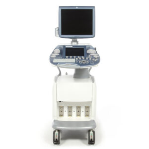
GE Voluson E8 Ultrasound Machine – Refurbished
MPIN: MP11239
Sign in to view priceAsk for Quote
Overview
Application training for the GE Voluson E8
KPI’s on-staff sonographer can provide onsite applications training or remote training via video conference for the Voluson E8 at a set price plus travel costs. A pre-recorded video training course is included in the sale, lease or rental of the Voluson E8 from KPI ultrasound.
Voluson E8 Service options:
Free technical support is available from KPI during installation and over the course of the standard limited warranty. Technical support is available after the warranty period at an hourly cost per issue.
GE Voluson E8 Maintenance:
KPI recommends the use of a surge protector along with a dedicated power outlet. Probes should be disinfected after every use with a disinfectant wipe proven not to damage the lens (KPI recommends SonoWipes for this.) One PM visit (preventative maintenance) every year.
GE Voluson E8 Dimensions & Weight
Height: (adjustable, maximum) 1393 mm (54.8 in)
Width: 580 mm (22.8 in)
Depth: 920 mm (36 in)
Weight: (no Peripherals) 131 kg (289 lb), approx 400 lbs with packaging
Voluson E8 Specifications
Digital Beam former
1,979,578 system processing channel technology
Displayed Imaging Depth: 0 – 30 cm
Minimum Depth of Field: 0 – 1 cm (Zoom, probe dependent)
Maximum Depth of Field: 0 – 36 cm (probe dependent)
Up to 274 dB Dynamic Range
GE Voluson E8 Electrical power
Voltage 100, 115-130, 220-240 VAC
Frequency 50/60 Hz (+/-2%)
Power Max. 1000 VA with on-board Peripherals
Thermal Output 3446 BTU/h
Review
GE Voluson E8 Review:
The GE Voluson E8 is most impressive when you are viewing it’s (HD live) high resolution 4D images. HD live is GE’s term for advanced 4D imaging in high definition. The resolution and detail of HD live versus traditional 4D is breathtaking. Think of when you first switched from a regular TV to an HD TV. Since GE launched HD live in 2012 several other manufacturers have created their own versions of HD 4D, but the Voluson E8 HD live is still the most popular. The detail, shading options, and fast frame rates are what keep GE in the lead for 4D. This extra 4D resolution gives Obstetricians a better volume assessment of the first trimester anatomy and also vascular structures. The resultant 4D images and movies can be exported in a variety of formats.
GE Voluson E8 Buyer’s Guide Awards:
Best Premium 3D/4D ultrasound machine 5 out of 5
Best Premium OB/GYN ultrasound machine 5 out of 5
Voluson E8 probe ratings:
The GE Voluson E8 is compatible with six matrix array transducers, including the amazing RM6C 4D convex, the RIC6-12-D 4D endocavitary, and the RM14L 4D linear. The Matrix array provides the highest resolution and best image quality available today. The E8 also offers a 50mm wide footprint linear probe specifically designed for breast imaging. The wider footprint allows scanning to be done in fewer passes. Strain based Elastography and Elastography Analysis (quantification analysis) is available for easy cyst detection and precise needle guided biopsies.
Voluson E8 ratings: standout features
SonoBiometry: A feature of the Voluson E8 that allows semi-automated biometry measurement for BPD, HC, AC, and FL.
SonoNT: Semi-automated, standardized measurements provided by the Voluson E8 of the nuchal translucency during the first trimester.
SonoIT: This is another semi-automated, standardized measurement on the GE Voluson E8 of intracranial translucency during the first trimester.
SonoAVC Follicle:This Voluson E8 feature gives extremely accurate automatic measurements of follicular volume. These are much faster and more accurate than taking manual 2D estimates.
SonoVCAD heart: A Voluson E8 computer aided display of the fetal heart from a 4D transducer that automatically generates a four-chamber view according to recommended standards.
SonoVCAD labor: automates the measurement of fetal head progression, rotation and direction during labor that is created by the Voluson E8 as an easy report.
Advanced STIC: STIC is Spatio-Temporal Image Correlation which on the Voluson E8 creates a full fetal heart examination in 2D and 4D including a view of the entire cardiovascular system.
Revisions
GE Voluson E8 BT10 to BT06 revisions
GE first launched the Voluson E8 expert in 2006 (15” LCD Monitor) . That first version was designated as BT06. “BT” is an abbreviation of “Break Through” and the number designates the year in which this version was launched. So the Voluson E8 expert BT08 (19” LCD Monitor) was launched in 2008 and was in production till the next version in 2009, the Voluson E8 BT09. The BT06 version had many problems with the software and hardware, which the BT08 version fixed. Version BT09 added SonoVCAD heart, Anatomical M-mode, and DICOM as standard features, and STIC, SonoAVC, and Scan Assistant as optional features. BT10 made Scan Assistant and STIC standard features instead of options, and added SonoRender Start, and SonoNT as standard features. Options on the Voluson E8 BT10 were SonoAVC, VOCAL, Advanced VCI and OmniView. The 3SP-D, and RRE5-10 probes were also added to BT10.
Voluson E8 BT12 and Voluson E8 BT13
The Voluson E8 BT12 added a new Dynamic Rendering Engine (new CPU & video card) to support HD live, GE’s high-resolution 4D in HD. The same options as BT10 were options with the Voluson E8 BT12, but BT12 also added support for the new C4-8-D, and RAB6-D 4D probes. The Voluson E8 BT12 really was a “BreakThrough” update! A smaller update came with the Voluson E8 BT13 that added SonoBiometry and Elastography Analysis (Elastography quantification). The new high-density imaging RIC6-12D is also available on the Voluson E8 BT13. The BT13 also have an additional feature such as V-SRI (Volume Speckle Reduction Imaging) on certain transducers. If you use the Flashlight V-SRI features while performing the HD Live on the Voluson E8 BT13, the 4D images are spectacular. That is the one of the main reason why GE Voluson E8 BT13 with HD Live is the most popular 4D imaging system in the world.
All revisions of the GE Voluson E8 expert
GE Voluson E8 (BT06)
GE Voluson E8 (BT08)
GE Voluson E8 (BT09)
GE Voluson E8 (BT10)
GE Voluson E8 (BT12)
GE Voluson E8 (BT13)
GE Voluson E8 (BT13.5)
GE Voluson E8 (BT14)
Popular configurations of the Voluson E8 in 2016:
- GE Voluson E8 (BT12 or BT13) with 4 transducers
RAB6-D 4D Convex
4C-D 2D Convex
IC5-9D 2D Endovaginal
11L-D 2D Linear
- GE Voluson E8 (BT12 or 13) with 1 transducer
RAB6-D 4D Convex
Probes
All GE Voluson E8 probes / transducers
4D Convex RAB2-5-D: [ 1 – 4 MHz ] 192 elements, 46mmR, max scanning depth 30cm
4D Convex RAB4-8-D: [ 2 – 8 MHz ] 192 elements, 46.8mmR, max scanning depth 26cm
4D Convex RAB6-D :[ 2 – 7 MHz ] 192 elements, 46.8mmR, max scanning depth 26cm
4D Convex matrix RM6C: [ 1 – 7 MHz ], 260 elements, Matrix Array 4D Convex (expensive)
4D Linear matrix RM14L :[ 4 – 14 MHz ], 960 elements, Matrix Array Linear (expensive)
4D Linear matrix RSM5-14: [ 5 – 13 MHz ] 960 elements, Matrix Array Linear (expensive)
4D Linear RSP6-16-D :[ 6 – 18 MHz ] 192 elements
4D Endocavitary multiplane: RRE6-10-D [ 4 – 9 MHz ] 192 elements, 11.7mmR
4D Endocavitary multiplane :RRE5-10-D [ 4 – 9 MHz ] 192 elements, 12.2mmR
4D Endocavitary matrix :RIC6-12-D [ 5 – 13 MHz ] 256 elements, 11.6mmR, max scanning depth 16cm
4D Endocavitary :RIC5-9-D [ 4 – 9 MHz ] 192 elements, 11.6mmR, max scanning depth 13cm
2D Endocavitary :IC5-9-D [ 4 – 9 MHz ] 192 elements, 10.8mmR, max scanning depth 16cm
4D Neonatal Pediatric Micro Convex: RNA5-9-D [ 3 – 9 MHz ] 192 elements, 15.4mmR
Convex 4C-D: [ 2 – 5 MHz ] 128 elements, 60.5mmR, max scanning depth 30cm
Convex: C1-5-D [ 2 – 5 MHz ] 192 elements, 56.1mmR, max scanning depth 30cm
Convex :C4-8-D [ 2 – 8 MHz ] 192 elements, 39.1mmR, max scanning depth 26cm
Convex :AB2-7-D [ 2 – 8 MHz ] 192 elements, 41.2mmR, max scanning depth 28cm
Convex :matrix M6C [ 1 – 7 MHz ] 960 elements, 50.7mmR, max scanning depth 26 cm
Linear :11L-D [ 4 – 10 MHz ] 192 elements, 37.4mm, max scanning depth 11cm
Linear :9L-D [ 3– 8 MHz ] 192 elements, 43mm, max scanning depth 14cm
Linear :matrix ML6-15-D [ 4 – 13 MHz ] 1008 elements, 49.6mm, max scanning depth 12cm
Linear :SP10-16-D [ 7 – 18 MHz ] 192 elements, 33.7mm, max scanning depth 6cm
Cardiac sector: 3S-D [ 1 – 3 MHz ] 64 elements adult cardiac sector
Cardiac sector :3Sp-D [ 1 – 5 MHz ] 64 elements adult cardiac sector
Cardiac sector :PA6-8-D [ 4 – 10 MHz ] 128 elements pediatric cardiac sector
Cardiac sector :S4-10-D [ 4 – 9 MHz ] 128 elements pediatric cardiac sector
Pedoff :(pencil probe) P2-D [ 2 MHz ]
Pedoff :(pencil probe) P6-D [ 6 MHz ]
Advanced Voluson E8 transducers: 4D and Matrix
The Voluson E8 BT13 Expert Series supports a wide range of 2D and 3D probes, enabling optimal quality images – especially in the first trimester and in complex gynecological exams. The RM6C 4Dconvex and RM14L 4D linear are next generation 4D volume matrix probes for high-resolution abdominal and vascular / musculoskeletal volume imaging that supports HD Live. The RM6C 4Dconvex and ML6-15-D linear probe features matrix technology for breast imaging, providing excellent spatial resolution and image uniformity in a 50 mm footprint that reduces the number of passes needed in a breast exam.
Popular GE Voluson E8 transducers
The RAB6-D 4D convex is a popular ultra lightweight 4D volume probe that is 40-50% lighter than the older RAB4-8-D convex, reducing user fatigue. The C4-8-D high frequency convex abdominal probe provides exceptionally high-resolution obstetric images from the first to the last trimester. The C1-5-D convex abdominal probe helps deliver a high level resolution and deep penetration, even on very large patients. The 9L-D wide-band linear abdominal probe helps provide high-quality images in the first trimester of pregnancy. The S4-10-D neonatal probe is dedicated to neonatology cardiac applications. The RIC6-12-D high-resolution 4D endovaginal probe helps detect fine details early in the first trimester and in gynecology exams.
Features
GE Voluson E8 Standard Features
19” LCD monitor on fully articulating arm
10.4” LCD touch panel
Automatic Tissue Optimization
Coded Harmonic Imaging with Pulse Inversion Technology
Coded Excitation (CE)
FFC – Focus Frequency Composite
HD-Flow
B-Flow
Virtual Convex and Wide Sector
Tissue Doppler
XTD (Extended View or Panoramic Imaging Mode)
SRI II (Speckle Reduction Imaging)
CrossXBeamCRI* (Compound Resolution Imaging)
TUI (Tomographic Ultrasound Imaging, aka: Multi Slice Imaging)
SonoNT
SonoIT
SonoBiometry
SonoRender Start
Scan Assistant
DICOM™ (Verify, Print, Store, Modality Worklist, Structured Reporting, MPPS, Media Exchange)
Static 3D Mode:
B Mode only
B + Power Doppler Mod
B + CFM Doppler Mod
B + HD-Flow Mod
B + CRI
Focus and Frequency Composite (FFC)
High Resolution Zoom
Pan Zoom
Steering
Virtual Convex
Wide Angle on endovaginal probes
BetaView
Patient information database
Image Archive on hard drive (via Raw-Data File , DICOM File both Single and Multiframe)
Image Archive to CD-R(W), DVD +/-
Export Images as BMP, TIFF, JPEG, AVI, MOV, DCM. MPEG4
3D/4D data compression (lossy/lossless)
Inversion
Real-time automatic Doppler calculations
Hard Drive 500 GB (BT13) , 450GB for data storage
OB Measurements Calculations & Reports
GYN Measurements Calculations & Reports
Vascular Measurements Calculations & Reports
Cardio Measurements Calculations & Reports
Abdominal Measurements Calculations & Reports
Multigestational Calculations
GE Voluson E8 technology definitions
Coded Excitation (CE) :This feature on the Voluson E8 improves image resolution and penetration in the far field. This allows use of a higher frequency on technically difficult patients.
Wide Sector :Standard on the Voluson E8, this widens the sector probe’s imaging field of view.
SRI II :A nonlinear diffusion filtering technique that improves image quality in real time by reducing speckles.
CrossXBeam CRI:This core technology on the GE Voluson E8 uses compound resolution imaging to improve border and image clarity.
TUI: This is a new visualization mode for 3D and 4D data sets on the Voluson E8. The data is presented as slices through the data set that are parallel to each other. An overview image, which is orthogonal to the parallel slices, shows which parts of the volume are displayed in the parallel planes. This method of visualization is consistent with the way other medical systems such as CT or MRI, present the data to the user. The distance between the different planes can be adjusted to the requirements of the given data set. In addition it is possible to set the number of planes. The planes and the overview image can also be printed to a DICOM printer, for easier comparison
of the ultrasound data with CT and/or MRI data.
SonoNT: This Voluson E8 feature allows for semi-automatic Nuchal Translucency measurements.
SonoIT: Sonography based Intracranial Translucency is a system supported measurement.Starting from the routinely used midsagittal view of the fetal face, obtained for assessment of the Nuchal Translucency and nasal bone, the ultrasound system uses a semiautomated mode to measure the anterior-posterior diameter of the fourth ventricle recognizable as intracranial translucency. The workflow is identical to SonoNT.
SonoBiometry: The Voluson E8 provides an alternative to the common fetal biometry measurements. It provides system suggested measurements for BPD, HC, AC and FL which need to be confirmed by the user or can be changed manually.
SonoRender Start: This feature on the Voluson E8 speeds up the acquisition of the fetal face in 4D.
Scan Assistant: Automated step-by-step help for those new to scanning.
Focus and Frequency Composite (FFC) :A Voluson E8 technology that utilizes two different transmission frequencies and two different focal ranges in the 2D image. This function combines a
low frequency to increase the penetration and higher frequency to keep a high resolution. It reduces speckle and artifacts in the 2D image to facilitate the examination of difficult-to-scan patients.
Beta View: allows the adjustment of the Volume O-Axis position of 3D probes in 2D mode. The green line in the displayed symbol indicates the position of the acoustic block. The “+” and “-“ define the corresponding sweep direction on the touchscreen.
Accessories
GE Voluson E8 Accessories
Sony UPD-897MD Digital Black & white thermal printer
Sony UPD-898MD Digital Black & white thermal printer
Sony UPX-898MD Digital Black & white thermal printer
Sony UPD-25MD Digital Color thermal printer
Mitsubishi P95DW Digital Black & white thermal printer
Mitsubishi CP30DW Digital Color thermal printer
Sony DVO-1000 DVD Recorder
CIVCO disposable biopsy guides (for Convex, Linear and Endo-cavity transducers)
Voluson E8 Supplies
Aquasonic ultrasound gel
Sono ultrasound wipes
Sony UPP-110HG thermal printing paper
Sony UPC-21L color thermal printing pack
Mitsubishi CK30L printing paper
Mitsubishi K95HG high gloss thermal printing paper
Voluson E8 ports
3 active transducer ports
1 inactive parking port
VGA Out
Network (RJ45)
Wireless Network interface (USB) (Option)
USB (6x external)
S-Video Out
DVI-D out
S-Video Out 2 (VTR)
S-Video In (VTR)
S-Video Out
Audio Out (Left & right channels)
Audio In (Left & right channels)
USB (5x internal)
RS 232: Optional, USB to RS232 converter
Parallel Port
Remote connections for external devices
Remote BW Printer via USB
Remote Color Printer/DVR via USB
Remote VCR (RS232) /DVR via USB
Remote Printer via Bluetooth Connection Kit (Option)
Footswitch via USB
ECG port
Options
GE Voluson E8 Options:
These are features that are not standard on the Voluson E8 BT13, but which can be added to the system for an additional cost.
Advanced 4D (inclusive of 4D Realtime, VCI, TUI, 4D Biopsy)
VOCAL II
Advanced VCI (Volume Contrast Imaging)
SonoVCAD-heart
SonoVCAD-labor
DICOM Connectivity
Elastography
Elastography Analysis
4D – Advanced STIC (Advanced Fetal Echo)
STIC (Spatio-Temporal Image Correlation, fetal heart analysis)
STIC + Power Doppler Mode
STIC + CFM Doppler Mode
STIC + HD-Flow Mode
STIC + CRI
STIC + CRI + CFM
STIC + CRI + PD
STIC + CRI + HD-Flow
STIC + B-Flow
STIC M-Mode
STICflow
SonoAVC (Follicles – Sono Automated Volume Count)
Coded Contrast Imaging† (not available in all countries)
Foot Switch, with programmable functionality
GE Voluson E8 options technology definitions
VOCAL II: A Voluson E8 option for cancer diagnosis, therapy planning and follow-up therapy control. It offers contour detection of structures and volume calculation. A virtual shell can be set around the contour of the lesion. VOCAL automatically calculates the vascularization within the shell by 3D color histogram by comparing the number of color voxels to the number of grayscale voxels.
Advanced VCI: (Volume Contrast Imaging) improves 4D contrast resolution. Volume Contrast Imaging utilizes the Voluson E8’s 4D transducers to automatically scan multiple adjacent slices and delivers a real-time display of the ROI. This image results from a special rendering mode consisting of texture and transparency information. VCI improves contrast resolution and therefore facilitates finding diffuse lesions in organs. VCI has more information (from multiple slices) and helps improve contrast due to improved signal to noise ratio.
SonoVCAD Heart: A Voluson E8 technology that automatically generates a number of views of the fetal heart to make diagnosis easier.
SonoVCAD labor: Allows the user to measure fetal progression during the second stage of labor such as fetal head progression, rotation and direction. Visual evidence and objective data of the labor process are provided. All SonoVCAD labor measurements are automatically added to the worksheet
Elastography: Visualization of the stiffness of tissue by assigning color values over B-mode.
Advanced STIC: An advanced Fetal Echo provided by the Voluson E8 that visualizes the fetal heart or an artery in static 3D.
SonoAVC follicle: A Voluson E8 option that automatically detects follicles in a volume of an organ (e.g., ovary) and analyzes their shape and volume. From the calculated volume an average diameter can be calculated. It also lists objects according to their size.
Coded Contrast Imaging: This option enhances the view of the Fallopian tubes vs. other tissue.
Imaging Modes
GE Voluson E8 Imaging Modes
B-mode (brightness)
M-mode (motion)
M-color flow mode
Anatomical M-mode
Trapezoidal mode
Color Doppler
Power Doppler
PW (Pulse Wave) Doppler
Bi-directional Power Doppler(HD Flow)
Steerable CW Doppler (Steerable continuous wave)
Tissue Doppler Imaging
Easy 3D (Freehand 3D)
Live 3D (4D)
HD Live (High resolution 4D) – only on BT12, BT13, BT14 systems
STIC (Spatio-Temporal Image Correlation)
B-Flow (Blood flow doppler)
Contrast Imaging
Strain-based Elastography
Applications
GE Voluson E8 Applications
Applications or Apps are the types of exams or studies that an ultrasound machine can do. More than this if an ultrasound machine supports a specific application it will have calculations, measurement and reporting software included to support those apps and make them useful in a clinical environment.
The GE Voluson E8 offers a broad selection of applications but is focused on OB/GYN and 4D.
4D Imaging
Obstetrics
Women’s Health(Gynecology & Breast)
Abdominal General Imaging
Fetal Echo
Strain Elastography
Small Parts (Breast, Thyroid, Testis)
MSK/Anesthesiology
Pediatrics
Urology (Renal, Prostate…)
Internal Medicine (Shared Service)
TCD (Transcranial Doppler)
Vascular
Contrast Imaging (GI)
Contrast Imaging (Cardiac)
Echocardiography Pediatric
Echocardiography Adult
Specification
System Overview
Year Launched : 2006
Estimated Market Price ($) : Premium
Monitor (inch) : 19″ LCD
Tilt/Rotate Adjustable Monitor : Yes
Monitor Resolution :
Image Size Resolution :
Touch Screen (Inch) : 10.4″
Trackball or Trackpad : Trackball
CP Back-Lighting : Yes
Weight : 265lbs(120kg)
Probe Ports : 3(+1 non active)
Battery : No
Boot-Up Time :
Sleep Mode (Quick Start) : No
Maximum Depth of Field : 0~30cm
Minimum Depth of Field : 0-2cm
Cart (HCU) : No
Independent Steer & Lockable Wheels : Yes
Imaging Modes
2D, M mode : Yes
M-color Flow Mode : Yes
Anatomical M-mode : Yes
Trapezoidal Mode : Yes
Color, Power Angio, Pulse Wave Doppler : Yes
Bi-directional Power (=HD FLOW) : Yes
SCW Doppler : Yes
Tissue Doppler(Velocity) Imaging : Yes
Freehand 3D : Yes
Live 3/4D OB/GYN : Yes
HD Live : Yes
STIC (Spatio-Temporal Image Correlation) : Yes
Live 3D Echo : No
Stress Echo : No
Strain and Strain Rate (Cardiac) : No
B Flow : Yes
Panoramic Imaging (=Logiq view) : No
Contrast Imaging – Cardiac : No
Contrast Imaging – General Imaging : Yes
Strain-based Elastography : Yes
Shear Wave Elastography : No
Features
Tissue Harmonic Imaging : Yes
Spatial Compounding(=CrossXbeam) : Yes
Speckle Reduction (=SRI) : Yes
Auto Image Opt(B mode) : Yes
Auto Image Opt(Doppler) : Yes
Write Zoom : Yes
Triplex Mode : Yes
Needle Enhancement or Needle Recognition : No
Auto NT Measurement (=Sono NT) : Yes
Auto Follicle 2D Measurement : Yes
Auto Follicle 3D Measurement : Yes(Semi)
Auto IMT : No
Auto IMT (Real Time) : No
Automated B/M/D Measurement : No
Automated LH Measurement(Automated Function Imaging(AFI), Cardiac Motion Quantification(CMQ), or Auto EF(Ejection Fraction) : No
Live Dual (B/BC) Mode : Yes
SmartExam or Scan Assistant : Yes
Fusion : No
Raw Data File : Yes
Flexible Report : Yes
Barcode Reader : No
Gel Warmer :No
Applications
Abdominal : Yes
Women’s Health Care (GYN & Breast) : Yes
OB : Yes
Fetal Echo : Yes
Vascular : Yes
TCD(Transcranial) : Yes
Small Parts (Breast, Thyroid, Testis…) : Yes
MSK/Anesthesiology : Yes
Pediatrics : Yes
Urology (Renal, Prostate…) : Yes
Echocardiography_Adult : Yes
Interventional Cardiology : No
Echocardiography_Pediatric : Yes
Echocardiography_Neonate : No
Stress Echocardiography : No
Transesophageal Echo_Adult : No
Transesophageal Echo_Pediatric : No
Internal Medicine w/ Shared Service : Yes
Surgury : No
Interventional Radiology : No
Contrast Imaging _ General Imaging (Low MI) : Yes
Contrast Imaging _ Cardiac (High or Low MI) : Yes
Bowel Imaging : No
Strain Elastography : Yes
Shear Wave Elastography : No
Transducers
Convex (1~6Mhz) : Yes
Convex (2~9Mhz) : No
Single Crystal Convex (1~6Mhz) : No
Single Crystal Convex (2~9Mhz) : No
2D Arrary 3D Convex (1~6Mhz) : No
Micro Convex (5~8Mhz) : No
Single Crystal Endocavity_Straight Type (3~10Mhz) : No
Endocavity_Curved Type (5~8Mhz) : Yes
3D Convex (2~6Mhz) : Yes
3D Convex Light Weight (2~7Mhz) : Yes(RAB6-D)
3D Endocavity (3~10Mhz) : Yes(6~12Mhz)
3D Micro Convex (3~9Mhz) : No
3D Linear (4~18Mhz) : Yes(14)
Linear (>14Mhz) : Yes(6~15Mhz)
Linear (3~12Mhz) : Yes
Linear (<9Mhz) : Yes
Single Crystal Linear (>14Mhz) : No
Single Crystal Linear (3~12Mhz) : No
Single Crystal Linear (<9Mhz) : No
Linear 50mm : Yes
Linear 25mm : No
Hockey stick (<13Mhz) : No
Hockey stick (>13Mhz) : No
T or L shape Intra Operative : No
Phased Array_Adult (1~5Mhz) : Yes
Single Crystal Phased Array_Adult (1~5Mhz) : No
2D Arrary 3D Phased Array (1~5Mhz) : No
Phased Array_Pediatric (3~8hz) : Yes
Single Crystal Phased Array_Pediatric (3~8hz) : No
Phased Array_Neonate (4~12Mhz) : Yes
ICE (Intracardiac Echo Cardiography) : Yes
TEE_Adult (3-7Mhz) : No
TEE_Pediatric (3~7Mhz) : No
2D Array 3D TEE (2~7Mhz) : No
Pencil CW (2Mhz) : Yes
Pencil CW (5 or 6Mhz) : Yes
Connectivity
DICOM 3.0 : Yes
DICOM SR_Cardiac : Yes
DICOM SR_Vascular : Yes
DICOM SR_OB/GYN : Yes
JPEG, WMV, & AVI : Yes
USB : Yes
HDD/SDD : 160GB
DVD/CD RW : Yes
Wireless LAN : Yes(Option)
Shipping Policy
Orders made at Medpick are initiated and processed for shipment upon receipt of request from the customer. Please note that our Shipping Services (Fee, Transportation, Loss or Damage of any shipment, etc.) are in accordance with the Seller\'s terms of Shipment.
Refund Policy
Please refer to Medpick Return Policy.
Cancellation / Return / Exchange Policy
Please refer to Medpick Return Policy.